appearance of red blood cells in Gram stains
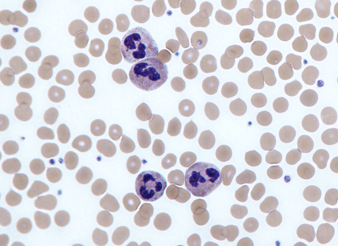
Red blood cells, white blood cells (polymorphonuclear neutrophils) and platelets
appearance of white blood cells in gram stains
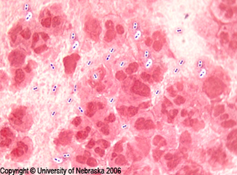
PMNs and bacteria
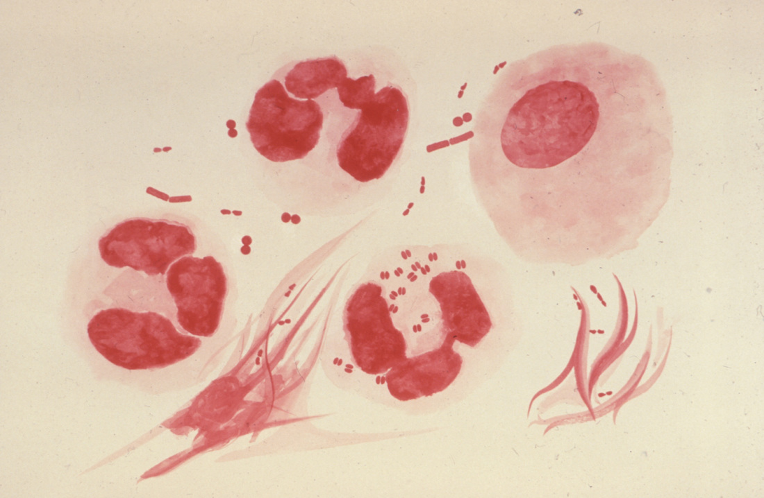
PMNs and bacteria
PMNs and bacteria

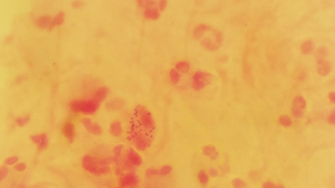
Appearance of squamous epithelial cells in gram stain
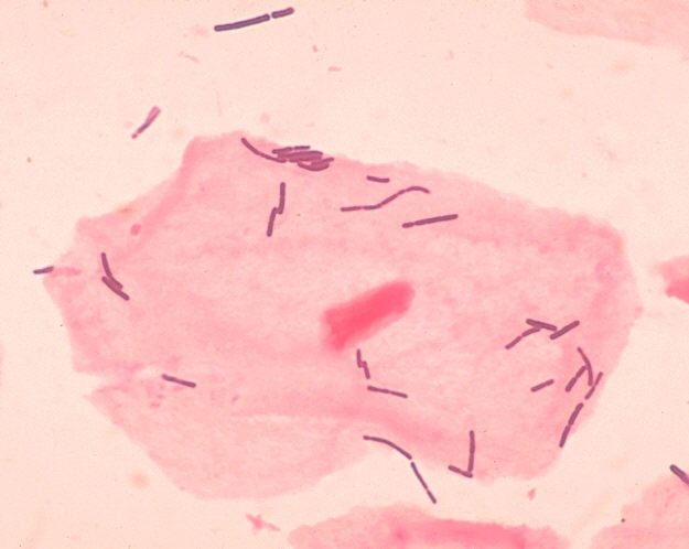
SEC and lactobacillus
